Core Facilities
Cryo-Electron Microscopy
On This Page
In This Section
Core Facilities
- General Information
- Animal-Based Studies
- Biochemical and Biophysical Studies
- Biorepository
- Cell-Based Studies
- Multiomic Analysis
- Informatics and Data Analysis
- Multiscale Imaging
- Pathology and Specimen Processing Resources
- Pharmacology
- Research Software and Applications
- Services
The Cryo-Electron Microscopy (cryo-EM) Core (RRID:SCR_021178) at the Penn State College of Medicine supports the modern demand for molecular imaging of both purified and cellular structures. It provides access to instruments for sample preparation as well as data collection. It is equipped with a Titan Krios (Thermo Fisher) cryo-transmission electron microscope that is fully equipped for automated data collection, a Hydra cryo-focused ion beam scanning electron microscope with high pressure freezing support and an LSM-980 (Zeiss) Cryo-Confocal microscope.
It was started in 2020 through the financial support of the Office for the Vice Dean of Research and the Pennsylvania Department of Health Tobacco Settlement Funds (CURE). Faculty directors Matt Swulius, PhD, and Kenneth Lee, PhD, have more than 20 years of combined experience in cryo-electron tomography and single particle analysis applications. They are available, along with facility manager Jennifer Sloppy, PhD, to provide guidance, training or data collection for researchers at the College of Medicine and elsewhere.
Instrumentation and Services
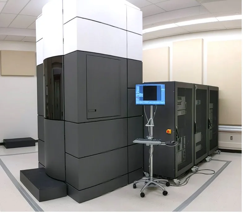
Titan Krios G3i
Titan Krios G3i (Thermo Fisher Scientific), 300 kV, equipped with Volta Phase Plates, Gatan Bioquantum energy filter and K3 direct electron detector (6k x 4k). This state-of-the-art cryo TEM is optimally equipped for the highest quality and highest throughput cryo-electron tomography and single particle data collection. This instrument is housed in room 149, NMR Research Facility, 30 Long Lane (Building 10 on the campus map).
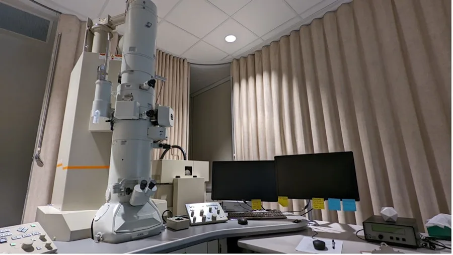
JEM 2100
JEM 2100 (JEOL), 200 kV, equipped with cryo box, Gatan side-entry cryo sample holder and Gatan Ultrascan CCD (4k x 4k). This microscope functions primarily as a screening microscope for working out initial freezing conditions prior to using the Krios for data collection. The JEOL 2100 is located in C1724, which is humidity controlled BSL2 laboratory space.
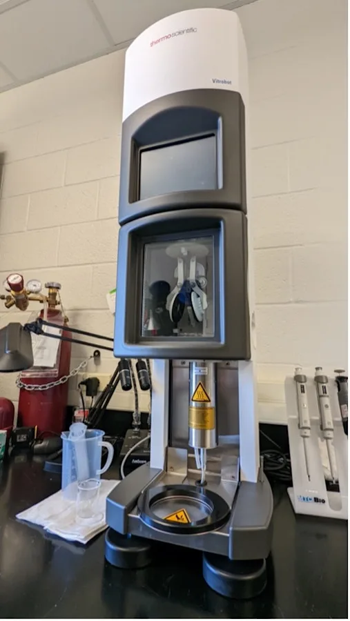
Vitrobot
The Mark IV Vitrobot standardizes and partially automates the vitrification process for cryo-TEM grids, and is available in C1724.
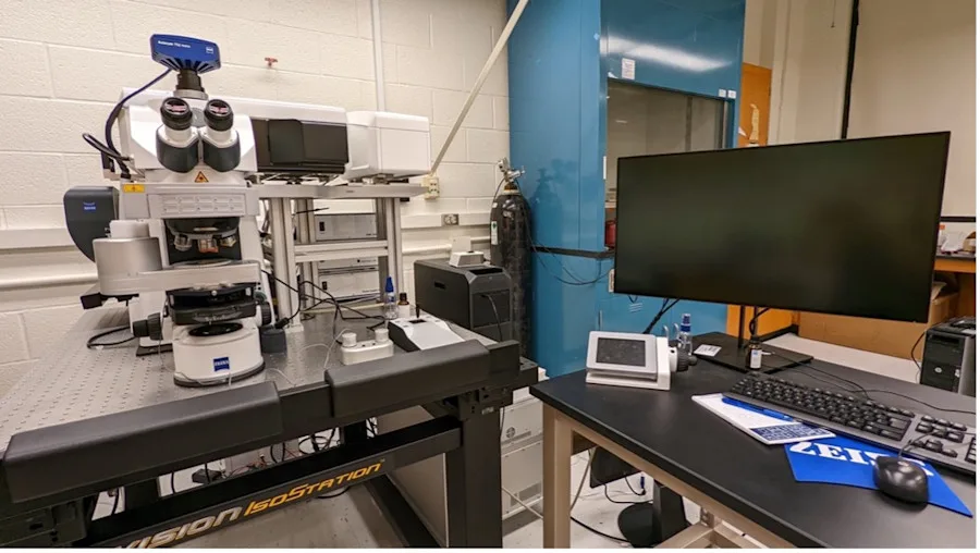
Cryo-Confocal
A Zeiss LSM980 Cryo Spectral Confocal microscope is available that is equipped with Linkam cryostage and a 34 Channel (2 MA-PMT and 32 Channel High Sensitivity GaAsP Array) detector array and an Airyscan 2 Super-Resolution High Sensitivity Detector with a 7 Laser System (405,445,488,514,561,594,639nm).
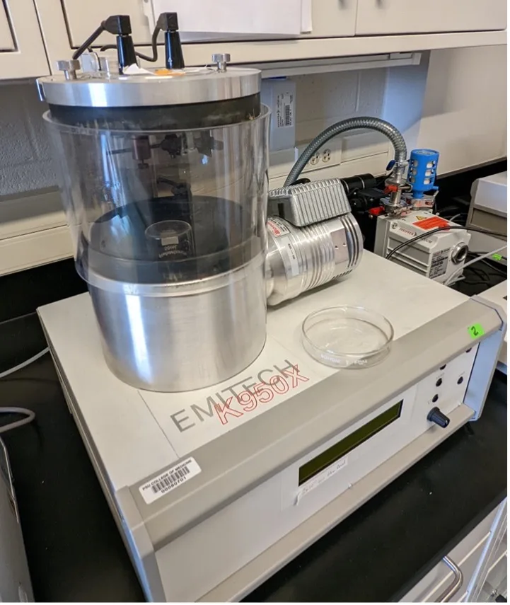
Sputter Coater/Glow Discharger
EmiTech High Vacuum sputter coater and glow discharge unit is available in C1724
Procedures, Protocols and Forms
General
This core provides services for Single Particle Data Collection, Tomographic Data collection and Cryo-Correlative Light and Electron Microscopy.
Training
Training is provided to users. Please contact the Core Manager at jsloppy@pennstatehealth.psu.edu.
Signups
Please register and use the iLab system.Go to iLab
Use
Users who are certified can use the facilities out of hours providing time is booked through iLab.
Shutdown and Cleanup
Certified users should follow the standard cleanup procedures detailed during their training. Failure to follow standard procedures might result in your access to this facility being limited.
Safety
If you are unclear about any process, please contact the facility manager at jsloppy@pennstatehealth.psu.edu.
Emergency
In case of an emergency follow the instructions you were given at the start of training.
Work With Cryo-EM
Fees
There is no charge for initial consultations and feasibility studies. For details on fees and options please contact Jennifer Sloppy at jsloppy@pennstatehealth.psu.edu.
Scheduling
Please contact Jennifer Sloppy at jsloppy@pennstatehealth.psu.edu.
Citation
All publications, press releases or other documents that result from the utilization of any Penn State College of Medicine Institutional Research Resources including funding, tools, services or support are required to credit the core facility and associated RRID for each core used. Use of an instruments in the cryoEM or cryoET cores should include the following:
“The CryoEM and CryoET Core (RRID:SCR_021178) services and instruments used in this project were funded, in part, by the Pennsylvania State University College of Medicine via the Office of the Vice Dean of Research and Graduate Students and the Pennsylvania Department of Health using Tobacco Settlement Funds (CURE). The content is solely the responsibility of the authors and does not necessarily represent the official views of the University or College of Medicine. The Pennsylvania Department of Health specifically disclaims responsibility for any analyses, interpretations or conclusions.”
Failure to do so might result in your access to services being impacted.
Contact the Core

Matthew T. Swulius, PhD
Associate Professor, Molecular and Precision Medicine

Jennifer Sloppy, PhD
Facilty support, Atomic Force Microscopy